Technology
Laterally Spreading Capsids

AAV.SPR ensures gene delivery
to large areas of the retina and safe delivery to the fovea

AAV.SPR, Atsena’s novel laterally spreading capsid, is the foundation of the company’s XLRS, USH1B, and Stargardt disease programs as well as our earlier discovery work. Developed to achieve therapeutic levels of gene expression in photoreceptors of the central retina while avoiding the surgical risks of foveal detachment, AAV.SPR’s benefits, the ability to spread beyond the subretinal injection site and safely deliver gene therapy to the fovea, have now been demonstrated in clinical trials for XLRS.
Atsena’s AAV.SPR capsid spreads laterally well beyond the margins of the subretinal injection bleb.

Credit: MoA animation by Visual Science, 2022

The preclinical studies below established the foundation for AAV.SPR’s clinical development. AAV.SPR has since been evaluated in patients with XLRS in the LIGHTHOUSE trial, where it demonstrated the ability to spread to the fovea, reverse structural damage to the retina, and improve visual function.


A preclinical study demonstrated that AAV.SPR exhibits enhanced lateral spread in subretinally injected macaques

AAV.SPR containing either myc-tagged human RS1 [AAV.SPR-RS1(myc)] or green fluorescent protein (AAV.SPR-GFP), both driven by a photoreceptor specific promoter, were co-delivered at a concentration of 6.6e11 vg/mL. AAV5 vectors containing identical constructs were used as a control.

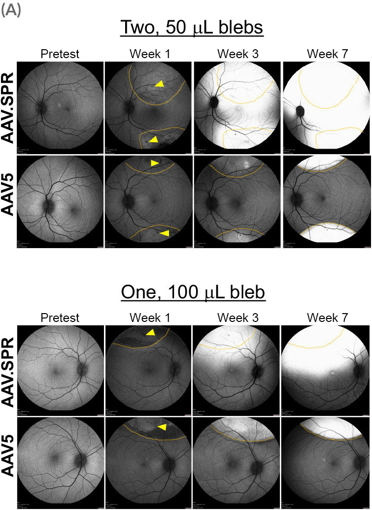
Eyes received either a single 100 µL bleb placed superior to the macula, or two 50 µL blebs placed superior and inferior to the macula. The location of injection blebs relative to the macula and GFP fluorescence over time were captured by confocal scanning laser ophthalmoscopy (cSLO). Borders of original blebs on day-of-dosing were confirmed by OCT and are outlined in yellow. AAV.SPR-mediated GFP expression was seen well beyond the margins of the injection blebs and expression increased over time. Foveal transduction was achieved following placement of either one or two injection blebs. In contrast, AAV5-mediated GFP remained confined to margins of the original bleb, and no foveal transduction was achieved.

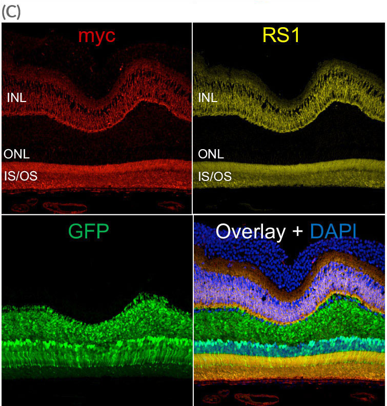
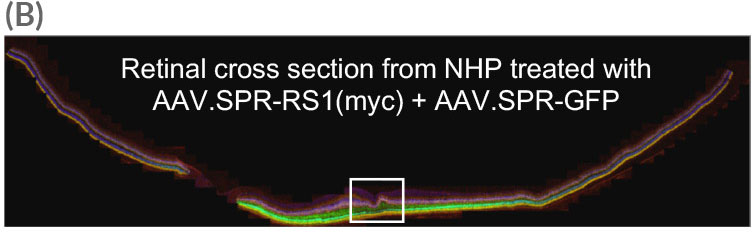
Widefield image of retinal cross section (taken from AAV.SPR treated eye) reveals widespread AAV.SPR-mediated GFP and myc expression. AAV.SPR-mediated GFP expression was found in the majority of foveal cones despite this region remaining fully attached during surgery. AAV.SPR-mediated myc (RS1) expression localized properly to photoreceptors and the INL, and colocalized with endogenous RS1 expression.

IS/OS = inner segments/outer segments of photoreceptors, ONL = outer nuclear layer, INL = inner nuclear layer


accessibility optionsAccessibility
Text Size
A
A